TECNIS Odyssey™ IOL
Hear From Your Peers
Precise vision. Every distance. Any lighting.
Enhanced Tolerance to Refractive Error†2
Optimized Dysphotopsia Profile†3
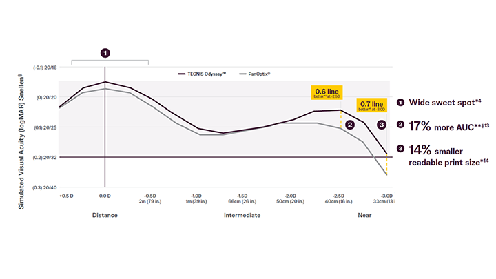
Unmatched Range of Vision**¶4
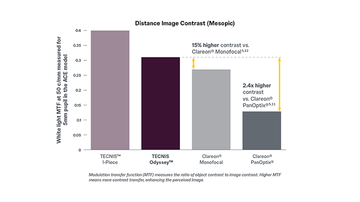
Best-in-Class Image Contrast^^†5,6,10
Optimized Dysphotopsia Profile
Freeform diffractive profile contributes to a low incidence of bothersome visual disturbances.9
93% reported no or mild halos, glare, or starbursts at one month post-op.9
Values rounded to the nearest 1%
Retrospective, multi-center, real-world clinical analysis of reported outcomes at 1 month post-operative visit, n=96. Symptoms reported without a specified severity level were classified as mild in the chart above.
Unmatched Range of Vision4,10
A continuous, full range of vision with better near compared to PanOptix®.*†

*Based on bench testing compared to PanOptix®
**AUC=Area under the curve
† continuous 20/25 or better
‡above 0.2 LogMar (~20/32 Snellen) compared to PanOptix®
§Snellen VA was converted from logMAR VA. A Snellen notation of 20/20-2 or better indicates a logMAR VA of 0.04 or better
Best-In-Class Image Contrast10
Superior low-light contrast compared to PanOptix®. *5,11,12

*Based on pre-clinical bench testing (white light MTF at 50 c/mm measured for 5mm pupil in the ACE model)
Best-in-class compares to competitor IOLs of comparable range of vision.
Learn More
With your consent, we will use your information to send you information about our products and services tailored to your interests through email. You may withdraw your consent at any time.
Please read our Privacy Policy.
This site is protected by reCAPTCHA and the Google Privacy Policy and Terms of Service apply.
*According to ISO 11979-7:2024, based on the clinical study of the parent IOL
¶Compared to PanOptix® based on bench testing and head-to-head clinical studies of parent lens
^^ Compared to PanOptix® based on bench testing (white light MFT at 50 c/mm measured for 3mm & 5mm pupil in the ACE model)
†Compared to TECNIS SYNERGY™ based on bench testing (white light MFT at 50 c/mm measured for 3mm and 5mm pupil in the ACE model)
**Continuous 20/25 or better based on pre-clinical bench testing for TECNIS Odyssey™
^ Values rounded to the nearest 1%. Based on 3-month postoperative data from a multicenter, observational clinical study in the U.S.
1. Data on File. 2024DOF4002 (prospective, multicenter, randomized, three-way-masked clinical study comparing subjects bilaterally implanted with TECNIS Synergy™ IOL (n=132) vs TECNIS™ 1-Piece Monofocal IOL (n=131) at 6-months post-op)
2. Data on File. 2024DOF4003
3. Data on File. 2024DOF4005
4. Data on File. DOF2023CT4023
5. Data on File. DOF2023CT4007
6. Data on File. DOF2019OTH4002
7. Data on File. 2024DOF4027 (ambispective, multicenter, observational clinical study evaluating subjects bilaterally implanted with TECNIS Odyssey™ IOL (n=33) at 3-months post-op)
8. Data on File. 2024DOF4029 (Based on 3-month postoperative data from a multicenter, observational clinical study in the U.S. evaluating visual and patient-reported outcomes from subjects bilaterally implanted with TECNIS Odyssey™ IOL (n=33))
9. Data on File. DOF2023CT4050 (Based on 1-month postoperative data from a multicenter, retrospective, real-world clinical study in the U.S. evaluating visual and patient-reported outcomes from subjects bilaterally implanted with TECNIS Odyssey™ IOL (n=96))
10. Dick, H. Burkhard MD, et. al. (2022 November). A Comparative Clinical Evaluation of a New TECNIS Presbyopia Correcting Intraocular Lens Against a Trifocal Intraocular Lens (“Forte 1”)
11. Data on File. 2024DOF4033
12. Data on File. DOF2018OTH4004
13. Data on File. 2024DOF4015
14. Data on File. DOF2023CT4056
INDICATIONS AND IMPORTANT SAFETY INFORMATION FOR TECNIS ODYSSEY™ IOL WITH TECNIS SIMPLICITY™ DELIVERY SYSTEM, MODEL DRN00V AND TECNIS ODYSSEY™ TORIC II IOL WITH TECNIS SIMPLICITY™ DELIVERY SYSTEM, MODELS DRT150, DRT225, DRT300, DRT375
Rx Only
INDICATIONS FOR USE
The TECNIS SIMPLICITY™ Delivery System is used to fold and assist in inserting the TECNIS Odyssey™ IOL, which is indicated for primary implantation for the visual correction of aphakia in adult patients, with less than 1 diopter of pre-existing corneal astigmatism, in whom a cataractous lens has been removed. The TECNIS SIMPLICITY™ Delivery System is used to fold and assist in inserting the TECNIS Odyssey™ Toric II IOLs that are indicated for primary implantation for the visual correction of aphakia and for reduction of refractive astigmatism in adult patients with greater than or equal to 1 diopter of preoperative corneal astigmatism, in whom a cataractous lens has been removed. Compared to an aspheric monofocal lens, the TECNIS Odyssey™ IOLs mitigate the effects of presbyopia by providing improved visual acuity at intermediate and near distances to reduce eyeglass wear, while maintaining comparable distance visual acuity. The lens is intended for capsular bag placement only.
PRECAUTIONS
1. This is a single use device. Do not resterilize the lens or the delivery system. Most sterilizers are not equipped to sterilize the soft acrylic material and the preloaded inserter material without producing undesirable side effects.
2. Do not store the device in direct sunlight or at a temperature under 41°F (5°C) or over 95°F (35°C).
3. Do not autoclave the delivery system.
4. Do not advance the lens unless ready for lens implantation.
5. The contents are sterile unless the package is opened or damaged.
6. The recommended temperature for implanting the lens is at least 63°F (17°C).
7. The use of Balanced Salt Solution or Ophthalmic Viscosurgical Devices (OVDs), is required when using the delivery system. For optimal performance when using OVD, use the HEALON™ family of OVDs. The use of balanced salt solution with additives has not been studied for this product.
8. Do not use if the delivery system has been dropped or if any part was inadvertently struck while outside the shipping box. The sterility of the delivery system and/ or the lens may have been compromised.
9. When performing refraction in patients implanted with the lens, interpret results with caution when using autorefractors or wavefront aberrometers that utilize infrared light, or when performing a duochrome test. Confirmation of refraction with maximum plus manifest refraction technique is strongly recommended.
10. The ability to perform some eye treatments (e.g., retinal photocoagulation) may be affected by the IOL optical design.
11. Recent contact lens usage may affect the patient’s refraction; therefore, in contact lens wearers, surgeons should establish corneal stability without contact lenses prior to determining IOL power.
12. The surgeon should target emmetropia as this lens is designed for optimum visual performance when emmetropia is achieved.
13. Care should be taken to achieve centration of the intraocular lens in the capsular bag.
14. Prior to surgery, the surgeon must inform prospective patients of the possible risks and benefits associated with the use of the device and provide them a copy of the patient information brochure.
15. Children under the age of 2 years are not suitable candidates for intraocular lenses.
16. The lens should not be placed in the ciliary sulcus.
17. Carefully remove all viscoelastic and do not over-inflate the capsular bag at the end of the case. Residual viscoelastic and/or over-inflation of the capsular bag may allow the lens to rotate, causing misalignment of the toric lens with the intended axis of placement.
18. The TECNIS™ Toric IOL Calculator includes a feature that accounts for posterior corneal astigmatism (PCA). The PCA is based on an algorithm that combines published literature (Koch, et al., 2012) and a retrospective analysis of data from a TECNIS™ Toric multi-center clinical study. The PCA algorithm for the selection of appropriate cylinder power and axis of implantation was not assessed in the prospective TECNIS™ Toric IOL U.S. IDE study and may yield results different from those in the TECNIS Odyssey™ Toric II IOL labeling. Please refer to the TECNIS™ Toric IOL Calculator user manual for more information.
19. The use of methods other than the TECNIS™ Toric IOL Calculator to select cylinder power and appropriate axis of implantation were not assessed in the TECNIS™ Toric IOL U.S. IDE study and may not yield similar results. Accurate keratometry and biometry, in addition to the use of the TECNIS™ Toric IOL Calculator (www.TecnisToricCalc.com) are recommended to achieve optimal visual outcomes for the toric lens.
20. All preoperative surgical parameters are important when choosing a toric lens for implantation, including preoperative keratometric cylinder (magnitude and axis), incision location, the surgeon's estimated surgically induced astigmatism (SIA) and biometry. Variability in any of the preoperative measurements can influence patient outcomes and the effectiveness of treating eyes with lower amounts of preoperative corneal astigmatism. The effectiveness of TECNIS Odyssey™ Toric II IOLs in reducing postoperative residual astigmatism in patients with preoperative corneal astigmatism <1.0 diopter has not been demonstrated.
21. Patients with a predicted postoperative astigmatism greater than 1.0 D may not be suitable candidates for implantation with the TECNIS Odyssey™ and TECNIS Odyssey™ Toric II IOLs, as they may not obtain the benefits of reduced spectacle wear or improved intermediate and near vision seen in patients with lower astigmatism.
22. All corneal incisions were placed temporally in the TECNIS™ Toric IOL U.S. IDE study. If the surgeon chooses to place the incision at a different location, outcomes may be different from those obtained for the TECNIS™ Toric IOL. Note that the TECNIS™ Toric IOL Calculator incorporates the surgeon’s estimated SIA and incision location when providing IOL options.
23. Do not reuse.
24. The safety and effectiveness of the TECNIS Odyssey™ IOL and the TECNIS Odyssey™ Toric II IOL have not been substantiated in patients under the age of 22 or those with preexisting ocular conditions and intraoperative complications, including those specified in the Warnings and Precautions, such as pupil abnormalities, prior corneal refractive or intraocular surgery, acute or chronic ophthalmic diseases or conditions (see below for examples).
Careful preoperative evaluation and sound clinical judgment should be used by the surgeon to decide the benefit/risk ratio before implanting a lens in a patient with one or more of these conditions.
Before Surgery
· Pupil abnormalities
· Prior corneal refractive or intraocular surgery
· Choroidal hemorrhage
· Chronic severe uveitis
· Concomitant severe eye disease
· Extremely shallow anterior chamber
· Medically uncontrolled glaucoma
· Microphthalmos
· Non-age-related cataract
· Proliferative diabetic retinopathy (severe)
· Severe corneal dystrophy
· Severe optic nerve atrophy
· Irregular corneal astigmatism
· Amblyopia · Macular disease
· Pregnancy
During Surgery
· Excessive vitreous loss
· Non-circular capsulotomy/capsulorhexis
· The presence of radial tears known or suspected at the time of surgery
· Situations in which the integrity of the circular capsulotomy/capsulorhexis cannot be confirmed by direct visualization
· Cataract extraction by techniques other than phacoemulsification or liquefaction
· Capsular rupture
· Significant anterior chamber hyphema
· Uncontrollable positive intraocular pressure
· Zonular damage
Potential complications generally associated with cataract surgery include, but are not limited to: endophthalmitis/intraocular infection, hypopyon, hyphema, IOL dislocation, persistent cystoid macular edema, pupillary block, retinal detachment/tear, persistent corneal stromal edema, persistent uveitis, persistent raised intraocular pressure (IOP) requiring treatment (e.g., AC tap), retained lens material, or toxic anterior segment syndrome, or any other adverse event that leads to permanent visual impairment or requires surgical or medical intervention to prevent permanent visual impairment. Adverse events that may be associated with use of the device include: IOL dislocation, tilt or decentration, visual symptoms requiring lens removal, residual refractive error, secondary surgical intervention (including IOL repositioning or removal).
25. Do not leave the lens in a folded position more than 10 minutes.
26. When the delivery system is used improperly, the lens may not be delivered properly, (i.e., haptics may be broken). Please refer to the specific instructions for use provided.
WARNINGS
1. Intraocular lenses may exacerbate an existing condition, may interfere with diagnosis or treatment of a condition or may pose an unreasonable risk to the eyesight of patients with:
a. Recurrent severe anterior or posterior segment inflammation of unknown etiology
b. Posterior segment diseases of which monitoring or treatment ability may be limited by an intraocular lens
c. Surgical difficulties at the time of cataract extraction and/or intraocular lens implantation that might increase the potential for complications (e.g., persistent bleeding, significant iris damage, uncontrolled positive pressure, or significant vitreous prolapse or loss)
d. Compromised posterior capsule or zonules due to previous trauma or developmental defect in which appropriate support of the IOL is not possible
e. Risk of damage to the endothelium during implantation
f. Suspected microbial infection
g. Congenital bilateral cataracts
h. Previous history of, or a predisposition to, retinal detachment
i. Potentially good vision in only one eye
j. Medically uncontrollable glaucoma
k. Corneal endothelial dystrophy
l. Proliferative diabetic retinopathy.
2. Patients should have well-defined visual needs and be informed of possible visual effects (such as a perception of halo, starbursts or glare around lights), which may be expected in nighttime or poor visibility conditions. Patients may perceive these visual effects as bothersome, which, on rare occasions, may be significant enough for the patient to request removal of the IOL.
3. A reduction in contrast sensitivity compared to an aspheric monofocal IOL may be experienced by some patients under certain conditions. The physician should carefully weigh the potential risks and benefits for each patient, with special consideration of potential visual problems before implanting the lens in patients including those with macular disease, amblyopia, corneal irregularities, or other ocular disease that may cause present or future reduction in acuity or contrast sensitivity, and should fully inform the patient of the potential for reduced contrast sensitivity and to exercise caution when driving at night or in poor visibility conditions after implantation.
4. Patients with a predicted postoperative residual astigmatism greater than 1.0 diopter, with or without a toric lens, may not fully benefit in terms of reducing spectacle wear.
5. Rotation of the toric lens from its intended axis can reduce its astigmatic correction. Misalignment greater than 30° may increase postoperative refractive cylinder. If necessary, lens repositioning should occur as early as possible prior to lens encapsulation.
6. Do not attempt to disassemble, modify or alter the delivery system or any of its components, as this can significantly affect the function and/or structural integrity of the design.
7. Do not use if the cartridge of the delivery system is cracked or split prior to implantation.
8. Do not implant the lens if the rod tip does not advance the lens or if it is jammed in the delivery system.
9. During initial lens advancement, quick advancement of the plunger is needed. Do not stop or reverse while advancing the plunger. Doing so may result in improper folding of the lens.
10. After initial lens advancement and the half turn rotation step, do not move the plunger forward until ready for lens implantation. Doing so may result in the lens being stuck in the cartridge.
11. The lens and delivery system should be discarded if the lens has been folded within the cartridge for more than 10 minutes. Not doing so may result in the lens being stuck in the cartridge.
12. Johnson & Johnson Surgical Vision, Inc. single-use medical devices are labeled with instructions for use and handling to minimize exposure to conditions which may compromise the product, patient, or the user. When used according to the directions for use, the delivery system minimizes the risk of infection and/or inflammation associated with contamination.
13. The reuse/resterilization/reprocessing of Johnson & Johnson Surgical Vision, Inc. single-use devices may result in physical damage to the medical device, failure of the medical device to perform as intended, and patient illness or injury due to infection, inflammation, and/or illness due to product contamination, transmission of infection, and lack of product sterility.
Third party trademarks are the property of their respective owners.
© Johnson & Johnson and its affiliates. All rights reserved.